Sunday, 16 February 2014

Beef production: Breeds and beef quality

Beef cattle are generally sturdier, meatier and larger than dairy breeds. In addition, they may have stronger maternal instincts, because in several systems the dam suckles her calves for several months. In dairy farming, calves are usually taken from the dam within 24 hours from birth. The information here is referenced from the website of Oklahoma State University and from other sources.

Angus or Aberdeen Angus is a medium-sized beef breed with either black or red coloring. Grows slower and gains fat faster than larger breeds.
Size (cows): 650-850 kg
Size (bulls): 1000-1300 kg


Belgian Blue has a mutation, which causes it to be double-muscled. The extreme size of its muscles causes severe problems when calving, and most cows must undergo several cesarean sections during their lives.
Size (cows): 700-850 kg (source)
Size (bulls): 1100-1250 kg

Blonde d'Aquitane, or blond, is a muscular and docile breed.
Size (cows): 700-900 kg
Size (bulls): 1200-1400 kg


Charolais is a white or cream-colored, large beef breed from France. It grows fast and generally gains fat slower than smaller breeds.
Size (cows): 700-950 kg
Size (bulls): 1200-1400 kg


(c) http://yallaroo.murrayfrancis.com/

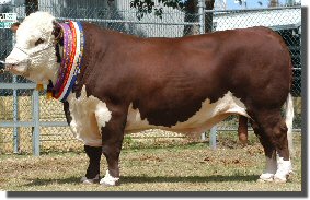
Herefords are massive, red and white colored animals. The head is usually entirely white and covered in curly, thick fur. Grows slower and gains fat faster than larger breeds.
Size (cows): 600-850 kg
Size (bulls): 1100-1300 kg


 Simmental is colored much like the Hereford, but the head is usually not entirely white. It is originally from the Simme Valley in Switzerland. It grows fast and generally gains fat slower than smaller breeds.
Size (cows): 700-950 kg
Size (bulls): 1200-1400 kg


Limousin  is another French beef cattle breed, and has originally been used as a working animal as well as for beef production. It has a high carcass percentage, i.e. the ratio between carcass weight and live weight. It grows fast and generally gains fat slower than smaller breeds.
Size (cows): 650-850 kg
Size (bulls): 1100-1300 kg


Carcass quality

Beef quality starts from carcass quality. A carcass is more valuable the less fat and bone is has, as the meat is the only economically important portion. The measurements are subjective, and based on the shape of the carcass. Most valuable cuts are evaluated especially carefully. In the European Union carcass quality is measured in an europ-scale: S > E > U > R > O > P (EEC  1208/81):

  • S (superior) = All profiles extremely convex; exceptional muscle development (double-muscled carcase type)
  • E (excellent) = All profiles convex to super-convex; exceptional muscle development
  • U (Very good) = Profiles on the whole convex; very good muscle development
  • R (Good) = Profiles on the whole straight; good muscle develop- ment
  • O (Fair) = Profiles straight to concave; average muscle develop- ment
  • P (Poor) = All profiles concave to very concave; poor muscle development
In addition to the EUROP-scale, the fatness of the carcass is evaluated from scale 1 (fat-free) to 5 (extremely fatty).

Two important concepts to consider are live weight and carcass weight. Live weight is the weight of the entire animal. Carcasss weight is live weight minus the weight of the head, genitalia, udder, digestive tract, internal organs, hide and hooves. The ratio between carcass weight and live weight is called carcass percentage. Carcass percentage varies between breeds, but is commonly 50-60 %.

When the carcass weight increases, to which the farmers often aim at, the relative proportion of lean meat decreases. In proportion, the amount of fat increases. The portion of the most valuable cuts (steak and filet) from the entire carcass does not change. Carcass weight can be increased by using plenty of concentrated feed, but a more effective method is to limit fattening by limiting energy intake at the finishing phase of the rearing.

Beef quality

Muscle becomes meat or beef after the animal has been slaughtered. Slaughtering causes chemical, physiological and biological changes in the muscle tissue. To prevent harmful changes, the animal and the meat must be handled correctly.

Beef quality consists of several factors:
  • Physiochemical properties: pH, color, sarcomere length, run-off, consistency
  • Chemical properties: Dry matter content, amount of protein, amount of fat
  • Sensory properties: juiciness, flavor, tenderness
Meat becomes stringy and chewy when the myocine and actin filaments of the muscle stick together after death (rigor mortis). Usually carcases are cooled under +7 C before rigor mortis sets in, which causes the muscle to constrict due to cold. To prevent cold constriction the meat may be stimulated with electricity. Once the meat is cooked, the proteins break and the meat becomes tender.

Juiciness means the amount of muscle fluid which is released when the cooked meat is bitten into. It
is related to the amount of fat in the meat, since fat increases the water retention capacity. The largest part, 64-80 %, of beef is water. The water is retained between actin and myocin filaments. Water retention capacity decreases as the pH decreases after slaughtering. The pH of muscle is 7,2, but it drops to 5,6 within 24 hours after death. Fat percentage varies between 2-25 %. 

If the animal has little glycogen in its body right prior to slaughter, the meat does not develop enough lactic acid after slaughtering and the pH does not drop as fast and low as it should. This results in a tar meat, or DFD meat (dark, firm, dry). DFD meat is not used for whole-meat products because it has poor shelf life. 


Friday, 14 February 2014

Beef production: Basics

 Beef production is the production of beef and veal, i.e. the meat from cows, steers, bulls, heifers and calves. First we look at the differences between beef production to the production of other types of meat. Then we discuss the anatomy of meat, the growth of the beef animals and their carcass composition. The different methods of rearing beef cattle are discussed in later posts.

What is beef? Beef is the meat from bovines, that is "cows" of different age, gender and breed. The beef you see in a market comes either from beef production, milk production or from a combined farm with both milk and beef production. In Northern Europe, for instance, nearly 90 % of all beed originates from the dairy industry.

Compared with other animals reared for their meat, cows are relatively inefficient at transforming vegetation into meat protein. Dairy cows produce much protein and energy to their milk, but as meat producers they are even less efficient than beef cows.The chart on the left shows only the energy and protein in edible cuts. Energy in tallow, lard etc is not included.

One can also compare the animal species in terms of how well they utilize nitrogen. Feed N recovery efficiency in the edible weight fraction is defined as the percentage of the N in the animal feed that ends up in the edible portion of the animal. N recovery efficiency is low in beef production, only 8 %, but much higher for pork (~20%) and poultry (~30%). (Oenema et al. 2005)

Growth models and carcass composition

Growth can mean either the actual daily growth of an animal, the extra growth it puts on due to management and feeding, or the increase in edible cuts. For example: A calf grows 1500 grams a day (actual growth). Of  that, 400 grams is due to heavy feeding (extra growth). After the calf is slaughtered one can then calculate backwards how much of the 1500g went to the edible meat (increase in edible cuts).

Growth consists of four types of changes:
  1. Changes in size (live weight)
  2. Changes in appearance (height, diameter of chest...)
  3. Changes in anatomical composition (fat percentage, ...)
  4. Changes in chemical composition (chemical composition of muscles, fat etc)
Change in size, for example the weight, is an inaccurate measurement because it is affected by what the animal has eaten and drank. The weight of the digestive tract of a bovine varies tens of kilos during the day. The weight of a cow inceases most rapidly from birth until 6 months of age, when the growth slows and finally comes to an end at the age of 2-3 years. The model is very simplified, because bones, muscles, connective tissue and nerves have a very different rate of growth. This also affects the third measurement: the anatomical composition. First the animal gains mostly bones and nerves, then muscles and finally fat, all altering the anatomical composition of the carcass.

Daily growth is measured either as the proportion of daily growth to the live weight, i.e. 1500g / 250kg = 0,006 %, or simply as g/day. Final weight of beef breeds varies from 350 kg (a Dexter cow) to 1400 kg (Charolais bull). Bulls grow 10-20 % faster than steers (castrated bulls), while steers grow as fast as heifers (Galbraith and Topps 1982).

Changes in appeareance only describe how the different parts of the animal grow in proportion to one another. For example a calf has tall feet and a shallow chest, while a grown cow has shorter feet and very wide chest.

Chemical changes describe the changes in the composition of the body. The higher the live weight, the more fat there is in a kilo of carcass, and therefore also the relative energy content increases. At the same time the relative portion of crude protein decreases. As with all young animals, first to grow are the bones and nerve tissue, with muscles next and finally body fat. Breed and gender also affect the carcass composition. Steers gain 10-45 % more fat than bulls, and breeds like Angus and Hereford are fatter than for example Limousin and Charolais. Note that breed does NOT affect the composition of the lean (fatless) carcass.

All in all, the growth of an animal is summarized in the picture below (Rumsey 1991).


The anatomy of muscles

30-40 % of the live weight of a bovine consist of skeletal muscles. The quality of edible meat is affected by the chemical, biochemical and physiological qualities of the muscle both before and after slaughter. Growth, feeding, animal handling and meat processing after slaughter also all have an impact on the quality of the meat.For bovines, 30 largest muscles contribute 75 % of the weight of all the muscles. The largest muscle groups are in the pelvic limb (hind quarters) with 28,5 % of live weight, and neck/thorax with 22,4 %.

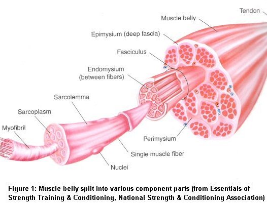
The anatomy of a skeletal muscle is shown in the picture to the left.The muscle is covered by epimysium, and consists of bundles of muscle fibres. The space between bundles is filled with perimysium, which has lots of nerves and blood vessels. Perimysium affects the tenderness of the meat. There is also fat between the muscle fibres, and this fat gives the meat it's marbling properties. Each muscle fibre is surrounded by endomysium, yet another type of membrane.


Each muscle fibre in the skeletal muscles has several nuclei. Fibres are surrounded by a sarcoplasm, which is a membrane, and sarcolemma, which is a type of elastic connective tissue. One fibre consists of 1000-2000 myofibrils. The functional unit of a myofibril is called a sarcomere. Sarcomere is where the muscle actually works, when thick and thin filaments of the sarcomere either slide closer or farther from a z disk (see the picture below). If the filaments become imbricated, the muscle constricts. When they slide farther apart, the muscle relaxes (returns to rest stage) or stretches. A very detailed video about the action potential and muscle activity can be found from Youtube.


Marbling and tenderness

Marbling of the meat means the increase of intramuscular fatty tissue, which occurs at the finishing phase of beef cattle rearing. Marbling is more pronounced with strong, grain-based feeding. The actual level of marbling is determined visually after slaughtering by estimating the percentage of fat in a cut of meat. Fat is seen as white areas in otherwise red meat.

The fat in the muscle is mostly based from de novo -fatty acid synthesis, which takes place in the rumen. The rumen biohydrogenates unsaturated fatty acids into saturated ones, so the fat of ruminating animals is more saturated than that of monogastric animals. For example, cattle have more saturated triglyceride 18:0 and less unsaturated 18:2 than pigs (Lawrence & Fowler: Growth of Farm Animals).

Tenderness is affected by the type of collagen in the perimysium, the connective tissue between bundles of muscle fibres. More important than types or the amount of collagen is cross-linking between the collagen types. Both the cross-linking and insolubility of collagen increase as the animal ages. That is why the meat from old animals is more stringent than the tender meat of young animals. However, when cooking meat in high temperatures even tender meat becomes stringent due to heat-induced chemical changes in the collagen.

Thursday, 16 January 2014

Managing risk in animal breeding schemes

Animal breeding is not exact science in the sense that normally one cannot exactly predict the outcome, or even select the "ingredients". Each gamete (an egg cell or a sperm) is different, and their combination and further cellular divisions all include an effect of randomness. So each breeding scheme has risks. This post will address some of those risks and how to minimize their impact.

Inbreeding

Breeding always requires some inbreeding. This is because we want to increase the genes from one or few excellent animals, so we use them for males/females of several generations. Consider horse racing and show jumping: it's common to list the famous parents, siblings, half-sibs and grandparents of any horse to prove its value.

The change of inbreeding can be calculated as
ΔF= 1 / 2Ne
where Ne is the effective population size. If the pnumber of parents of different sexes isn't equal, then we estimate

ΔF ≈ (1 / 8Nm) + (1 / 8Nf)

Inbreeding works in two ways: inbreeding increases variance between lines/populations, but decreases variance among a line/population. Remember that inbreeding depression, the negative effect of inbreeding on genetic diversity, can be negated by breeding two animals of completely different lines.

Genomic selection versus progeny testing

Both agenomic selection schemes (GS) and progeny testing schemes (PT) have their own risks. Professors Alban Bucket  and Jarmo Juga from the University of Helsinki have studied the risks in bovines. They state that in GS schemes the rate of inbreeding is slightly higher than in PT schemes, but reciprocally the genetic response is much higher in GS than in PT. The choice becomes a matter of balancing the risks. How high of an inbreeding level do we accept to get strong genetic response? 

Bucket and Juga state that if the amount of sires is not increased, the risk is comparable between GS and PT schemes. The risk in GS can be further minimized by increasing the amount of MOET (multiple ovulation, embryo transfer) and the number of genotyped females.This increases the genetic diversity and allows effective Mendelian variance. However, increasing the amount of AI bulls in a GS scheme increases the risks of inbreeding.

Preserving genetic diversity

As has been stated earlier, selection and inbreeding impact genetic diversity in two ways: the variance between lines increases, while the variance within lines decreases. If a line equals a breed, the impact can be very strong.One example can be found from the study by Uimari and Tapio, who studied how the effective population size has changed over generations in two pig breeds. During 50 generations, selection has decreased the effective population size from 600 to a mere 50. The decrease is simply due to breeding selection.

The impact of selection to the Ne of two pig breeds.
(c) Uimari and Tapio

Maintaining genetic diversity should be duly considered in every breeding scheme. By genotyping a large amount of animals it is possible to ensure diversity by pairing unrelated animals. By genotyping one can also ensure that rare alleles stay in the population, and that there is enough heterozygozity. These two go often hand in hand: rare alleles are found most often in heterozygotes than in homozygotes. By genotyping one can also preserve traits of specific interest and genomically control the level of inbreeding.

FAO, The Food and Agriculture Organization, has created a simple chart about preserving genetic diversity. The chart is part of their publication considering The State of the Worlds ANGR for Food and Agriculture (ANGR = Animal genetic resources). It shows that the actions required are rather simple. Because really - 
all it takes is the courage to look beyond monetary gain and efficiency.


Monday, 13 January 2014

Calculating breeding values

Basics of animal breeding have been covered earlier in this blog. We've discussed the very basics of animal breeding as well as the  Mathematics of animal breeding . Optimization of animal breeding schemes has also been briefly considered. Today we take a closer look at calculating the breeding value using statistical concepts and information from various sources.

Estimated Breeding Value


When calculating EBV (estimated breeding value) for an animal, we usually want to combine information from various sources. We have results from the animal itself, but also from its relatives. However, EBV is always for one trait only.

The formula for an EBV is

 = b1x1 + b2x2 + ... bnxn

where  is the EBV, b1 is the regression coefficient for trait 1 and x1 is the result for trait 1. Seems simple, doesn't it? Now all we need to do is calculate the b values. The key to the b values is to remember where dealing with several bits of information at once, and every "bit" is actually an equation

 Â = (y1 - μ) = a1 + e1

where  is the EBV, y1 is the animal's own result in trait 1, μ is the population mean result in trait 1, a1 is the additive genetic effects contributing to the trait and e denotes the environmental factors contributing to the trait. Simply put: an EBV consists of genetics and environmental factors.

So, the b's must fulfill equations for traits 1 ... n at the same time. Instead of a group of equations we use matrices to calculate the b's. Only then can we continue to calculating the actual selection index. The matrix notation for calculating b's is
b = P-1G

Here P and G are matrices. P includes the variances and covariances between phenotypic results. The marking P-1 simply means that the transpose of the matrix P is used in the calculation. G is another matrix, which links the information sources to true breeding values. In the G matrix Ai denotes the true breeding value of animal i. Info1 and Info2 below are different information sources, for example animals 1 and 2.


You might remember that we never know the actual breeding value A, so we always work with the estimated BV, denoted as Â. However, with enough information and correct calculations it is assumed that A = Â. The trick in matrix G is to consider the genetic relationships between the information sources, here animals 1 and 2. If Info 1 is the animal itself, so that the source for info 1 = i, then Cov(Info 1, Ai) = Var(Ai). More generally,

Cov(Info x, Ay) = a(x,y) * Var(A)

and

Var(A) = h2 * s.d. (P)

where a(x,y) is the coefficient of genetic relationship between animals x and y. If x and y are full siblings, their coefficient of genetic relationship is 0,5, and Cov(Info x, Ay) = 0,5 Var(A). If they're half-sibs, it's Cov(Info x, Ay) = 0,25 Var(A) and so on. s.d. (P) is the standard deviation of the trait P, or the trait for which we are calculating the breeding value for. Standard deviation of P is the square root of the variance of P.

Selection index and economic breeding value


So now we can calculate the EBV for one trait. What if we want to combine several traits into one number? Then we need a selection index. It works like EBV, but combines information from several sources and several traits into one.


A selection index can either be optimal or common. The difference is in the coefficients: optimal coefficients minimize the variance between true breeding values and estimated breeding values. In a common selection index the coefficient b is said to be "any b0", but in the optimal index b = P-1Cv. The optimal index considers covariances, and breeding accuracies impact the b coefficients.

Here we can see a new matrix, C. C is used if the measured traits are not the same as the traits to be improved. For example, we might measure weight and thickness of back fat, but we want to improve weights and percentage of lean meat. Now we need the matrix C, which relates to the other matrices as shown in the picture below.


If we want to include money to the calculations, we get a total breeding value. Money is used in breeding values to give economical weights to each trait. This weight is entirely decided by animal breeders, and based on what they think is most important. Economical weight isn't linked to genetics or phenotype in any way. It is simply a way to put the traits into some order of importance.Often economical values is derived from actual profits or costs regarding the trait in question. The weight is currency per 1 unit of increase/decrease in the trait, for example euros per +- 1 kg of meat or dollars per +- 1 weaned piglet. The economical value can be used to ompare the costs and profits between different breeding schemes.

(c) Wikipedia Commons
For example:
We have two schemes for pig breeding. One scheme gives us - 0,5 piglets per sow, but 10 kg more meat since the surviving piglets are heavier. The other scheme gives + 0,7 piglets but -6 kg meat.  Let us assume that 1 kg of meat is +10 euros and 1 piglet is 15 euros.

Now the first scheme yields (-0,5 * 15) + (10*10) = 92,5 euros, and the second scheme (0,7 * 15) + (-6 * 10) = -49,5 euros. With these exaggerated numbers it is easy to see which scheme would be more profitable for the producer.

Economic weight can also be used when restricting a selection index. We may want to improve one trait, but leave another trait untouched. In that case the economic value of the trait, which is not allowed to change, is set to 0,

Total breeding value


Using indices and economic breeding values we can calculate a total breeding value for an animal. The formulas are

H = v'g
I = b'x where b = P-1Gv

 H = total breeding value, estimated using the index I
v = economical weights of traits
g = breeding values of traits
I = selection index
b = regression coefficients for traits
x = vector of observations [result1    result2    resultn]
P = covariances and variances between observations
G = covariances and variances between traits to be improved and measured traits.

Additional information and sources

Mrode, R. A. Linear Models for the Prediction of Animal Breeding Values, 2nd edition. CABI Publishing, USA. ISBN-13: 978-085199-000-2

Cameron, N. D. Selection Indices and Prediction of Genetic Merit in Animal Breeding. CAB International, USA. ISBN-13: 978-085199-169-6

GenUp-software for playing with genetics: http://www-personal.une.edu.au/~bkinghor/genup.htm

Monday, 9 December 2013

Fur farming - breeding and welfare

When talking about animal breeding, it is important to understand what it is. Breeding aims at improving the genetics of an animal population with methological breeding systems and programs. Breeding has clear targets, traits to be measured, recorded and followed etc. Breeding is not about increasing the animal population by mating animals to one another, which is what some pet "breeders" do.

Colors of mink pelts (c) Fur Commission
Fur animal breeding has always aimed at producing pelts which fetch the highest price in the market. Three main qualities affect the price: the size, quality and color of the pelt. Size is measured as the length of the pelt. As a breeding target larger pelts require larger animals, so often the largest and the ones that grow fastest are selected as breeding animals. Large in this context means fat. This however has lead to severe problems with fertility: fat females give birth to small litters. Fur animals breeders must therefore balance between size and litter size to produce enough pelts of an adequate size.

Pelt quality consists of several traits. The most important quality factors are
  • flaws (such as bite marks)
  • the quality of the guard hairs
  • the quality of the undercoat
The quality of the pelt is a combination of its mass and the coverage of the guard hairs. The ratio between guard hairs and undercoat contributes to the mass of the pelt. In a pelt of good mass the undercoat is thick, strong and elastic, and it supports the guard hairs. In a high quality pelt the guard hairs are longer than the undercoat, there are no color flaws and the length of the hairs is even.

The third important quality factor is the color of the pelt. The desirability of different colors varies yearly and depends on fashion trends. Color can be determined by several qualities, such as hue and darkness.

Breeding traits

The breeding objectives can be divided to three classes, all of which may or may not be used for all fur animal species. The targets are evaluated in a two-step process, first in grading and then as pelt quality factors.

Grading is a process where the quality and color of the fur of live animals is manually estimated. Four qualities are estimated: the size, color, purity and quality of the pelt. Grading is done at the fur farm, and may be done several times a year. The size of the pelt is either estimated or measured. Pelt size is measured from the tip of the nose to the beginning of the tail. The purity of the color is also evaluated. For blue foxes the pelt can have four hues ranging from blue to red. The darkness of the color is graded and the mass of the pelt is estimated.

Mink pelts at an auction (c) Searching for Style
The same traits are measured again after skinning in a process called evaluation of the pelt quality. The differences are that pelt quality evaluation is done post-mortem and usually automatically, while grading is done when the fur isn't yet fully developed and is a manual task. The number of classes to which pelts are classified also varies between grading and pelt quality evaluation. For example in grading the pelt quality gets a score from 1-5, while the pelt quality classes depend on the company selling the pelts.  There are six traits evaluated as pelt quality: grading qualities (the size, color, purity and quality of the pelt) plus mass and coverage of guard hair.


Fertility is probably the most important breeding target. There are two main fertility traits:
  • Litter size = number of puppies alive at 3 weeks of age / number of dams with at least one 3wk old pup
  • Litter result = number of puppies alive at 3 weeks of age / number of mated females
The litter size and result are counted after the pups are three weeks old, because the highest pup mortality is during the first weeks of life. Studies show that young blue fox dams produce larger litters, but the pups have higher mortality than the pups of older females. 2 year old blue fox vixens have the best litter results.

Heritabilities (h2) and genetic correlations in blue foxes

Heritabilities are a way of measuring how much genes impact a certain trait, i.e. how well can the trait be developed by animal breeding. Traits with high heritability are easier to develop than traits with very low heritability. Heritability ranges from 0 to 1, where 0 means that genes have nothing to do with the trait, and 1 means that the trait is affected by genes only and there's no environmental impact at all.

A Finnish doctor of animal science, Jussi Peura, has calculated heritabilities to several breeding traits for the blue foxes. He found that pelt traits have the highest heritabilities and fertility traits have the lowest. For example, color darkness has a heritability of 0.55 and pelt size 0.30. On the other hand, litter size at 1st parity (1st litter) had a heritability of 0,1, which is fairy low.

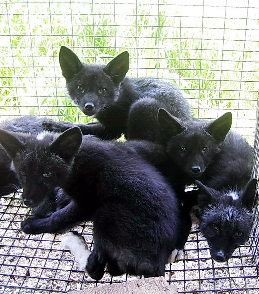
Litter of silver foxes (c) Bioacoustica
Peura also studies genetic correlations, i.e. how much traits depend on other traits. For example, in humans height often correlates with weight: the taller, the heavier. For blue foxes there were clear positive genetic correlations between size in grading and size in pelt quality (0,74), color in grading and pelt quality (0,84) and quality in pelt quality and mass in grading (0,75). When a correlation is positive, both traits increase simultaneously.

Negative correlations mean that increasing one trait decreases the other. Blue foxes had a significant negative correlation between size and litter size (-0,28). There were mild negative correlations also between size and color purity in grading (-0,25) and color in pelt quality and size in grading (-0,17).

What all these mean is that grading gives a reliable estimation of the actual size and quality of the pelt. However, the purity estimation in grading is a poor estimation of the actual purity. The correlations also clearly show that increasing the size of the animals result in poor litter results.

Welfare of fur animals

The issues and solutions presented in this text are based on WelFur, which is again based on WelfareQuality -protocol. Welfare issues here are classified under the four basic principles of WelfareQuality: good feeding, good housing, good health and appropriate behavior.

Animal welfare is a complicated concept with several different definitions and theories. Here animal welfare means the subjective experience the animal has about its own psychological and physiological state as the animal tries to adapt to its surroundings. Welfare cannot then be measured directly. Animal cannot have a welfare of "9.5" or "good" - we can merely measure its behavior and surroundings, and deduce the level of welfare from the findings.

Good feeding


Body condition score for blue foxes
As has been previously discussed, obesity is a severe problem for blue foxes. They naturally would eat a lot during fall, and show this behavior also in fur farms. In farms feed is easily available and high in energy, so the animals gain weight very fast, and the size of their pelt increases. Obesity causes their front paws to bend, which again makes mobility very difficult. Healthy paws are rare in any blue fox farm.

Fat animals cannot breed well, so the animals kept alive for breeding are nearly starved during the winter. While it would be natural for them to lose weight, in farms the difference between the Fall weight and Spring weight is much greater than in the wild. Normally a blue fox would weigh 3-5 kg - the average weight of farmed blue fox males is a staggering 19 kg, and 10 kg for the females.

In any farm where animals are kept in group cages there is no peace during feeding. The animals fight over the food, which causes stress, and fearful animals may be underfed while the more dominating animals are overfed.

Another problem common to all fur farms is the availability of fresh, clean water. During winter the water pipes may freeze, and during summer the water may heat and become unsanitary. Problems with availability of water are usually technical in nature: unlike with feeding, there is no reason to purposefully limit the animals' intake of water.

Good housing


Good housing is a wide concept, which consists of a comfortable place to lie, warmth and the easiness of mobility.

Foxes on a shelf (c) Dyrevern Alliansen
Small fur animals, like minks and ferrets, must have a nest box available all year round. Foxes have a nest only during whelping. The nest is the only part of the cage with solid floor and walls: otherwise the animals sleep, play and walk on a metal net ( = mesh). The nest box has been proven to increase fearfulness towards humans, because it gives a place for the animals to hide to. That, and the animals' tendency to defecate on solid surfaces, are the main reasons for keeping the nest box available only for a short while. 

Foxes must also have a shelf in their cage, unless they can lie on top of the nest.The shelf is actually very important for the foxes, who prefer high places from where they can scan their surroundings.

The main reason for using mesh flooring is that urine and feces pass through it. Fur animals have a tendency to defecate on a solid surface, which would then need to be cleaned daily to prevent the animals from soiling their fur. While a mesh floor sounds unpleasant, foxes actually prefer mesh over earth floor or solid floor such as wood at least as a resting place. This may be because the mesh allows their fur to stay "puffy" so the animal stays warm. There are no similar studies done on other fur animal species.

Good health

In order to have and to maintain good health, the animals must have good housing and good feeding. Good health means that the animals have no sicknesses, but also no internal or external injuries or disabilities. In fur farms mortality due to diseases is somewhat low, 3-4 % in foxes between April - October. Treatment of illnesses may be rare. Many animals are kept until skinning even if they are sick, or left to die.


Disease epidemics in fur farms are relatively rare. The only exception is plasmasytosis for minks, which occasionally causes significant losses due to sickness and exterminations. The most common diseases vary between animal species. For foxes and minks infections of the womb and gut are the most common illnesses. Eye and skin infections, urinary tract infections and diarrhea are sometimes seen in foxes. Diarrhea can also infect minks, but rarely raccoon dogs, who are capable of eating even partially rotten meat without trouble.

As has been noted before, bent front paws and obesity are extremely common health risks in blue foxes. They are not usually life-threatening or even painful in fur farm conditions, but most likely they do decrease the animal's welfare.

Compared to most farm animals (cows, pigs etc.) fur animals receive no painful treatments. For example, there are no surgical castrations, tooth cutting or cutting of the ears, which are all performed on piglets. Still, care must be taken to ensure that all fur animals are killed humanely and quickly before skinning. The killing and skinning must be done far away from the live animals to prevent fear and panic.

 Appropriate behavior

Expressing normal and appropriate behavior is mostly impossible in fur farms. Pups can be weaned at the "correct" age, but they cannot spread apart like they would do in the wild. Animals are not allowed a space for their own. They also do not need to hunt or forage, which in the wild would occupy most of their time.

Silver foxes would normally live in small groups, where only the dominant female would raise a litter, and the females of lower rank would kill their own pups if they have any. To keep thousands of females in adjacent cages, all with their own litter, may cause stress and increase situations where females kill their pups.

Group vs single housing is an important question for all fur animals, but many study results are contradictory. Group housed minks can fight more and therefore damage the pelts. Their stress levels may also be elevated if the group is not balanced. On the other hand, group housed minks have company, can play with one another, and in a balanced group are also less stressed than individually housed animals.

Tuesday, 3 December 2013

Fur farming - animal species and annual cycle

Fur animals are farmed to provide raw material for clothes designers and the fashion industry. The ethics of fur farming is an important topic and should always be considered, but it will not be covered here. This post is about the practices of fur farming, fur animal species and caring for fur animals.

Every year nearly 60 000 000 mink pelts and 4 000 000 fox pelts are produced, with Denmark as the leading producer of mink pelts (over 15 million annually) and Finland of fox pelts (nearly 2 million). When these numbers are considered, it is obvious that fur farming is important to the national economy to some countries. Still, major fur producing countries like the Netherlands have banned fur farming due to ethical concerns. Finland's house of parliament voted on the subject in 2012, and vetoed a bill demanding to end fur farming.

Fur animal species

Mink (Neovison vison)
Minks are the smallest of fur animals, weighing 1-4 kgs and ranging from black to brown to white in color. Minks, like all fur animal species, come to heat once in a year in the spring. Female minks are in heat during the beginning of March. Mating induced ovulation, so the mating time does not need to be carefully planned, and usually each female is mated 2-3 times. One male is allowed to mate with 4-5 females.

The gestation period for minks is 40-70 days. Most minks in one farm have their litter within 2 weeks from each other. Naturally minks would deliver 6-7 pups, but in captivity only 4-5 pups survive to adulthood. Minks are killed with gas (CO / CO2) and skinned in November, apart from breeding animals which are kept over the winter and mated again in Spring.

Minks are raised either alone, or two or four animals in one cage. Studies show that group housing reduces stereotypical behavior, but only if the groups are kept steady and balanced. Usually groups consist of pups from the same litter, with one female and one male, or two of both. In Europe, four pups or 2 adult animals may be kept in a cage of 2550cm2 in size. In the wild each mink would live alone in a territory covering several hectares.

(c) greengirlabroad
Blue fox (Vulpes lagopus)
The blue fox originates from the endangered arctic fox. Its weight ranges from 8-12 kg, but the largest males can be near 19 kgs. The largest animals are not muscular but obese: foxes are overfed to make them fat, which increases the size, and thus the price, of the pelt.

Obese animals suffer from major health issues, such as bent legs and difficulty to move. In recent studies in Finland, blue foxes with healthy legs are a rarity. Because fat animals breed poorly, the animals kept for breeding are kept on minimum food during the winter, so they lose weight and are able to breed in the Spring. While it mimics the natural habit of the animals (gathering body fat in the fall for the harsh winter), in farms it is taken into extremes and thus causes major stress for the animals.

Blue fox females, vixens, are in heat for 4-5 days during February and March. Most vixens are artificially inseminated using sperm collected from male foxes in the same farm. The gestation lasts 51-53 days, and each litter has 5-7 pups. This is a very poor result compared to the litter size of wild arctic foxes, which is 8-10 pups.

Blue foxes have a very thick fur to keep them warm. Because on solid surface the fur would flatten, the animals actually prefer net flooring to solid flooring. Still, overgrown claws and wounded paws may occur even if the net is covered with plastic.

(c) Wikimedia commons
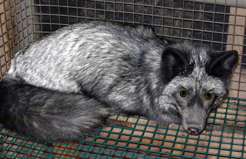
Silver fox (Vulpes vulpes)
Silver foxes are descendants of the common red fox. They are smaller and leaner compared to the blue fox, and their fur is less thick. Silver foxes are not fattened and then starved like blue foxes, because due to their heritage they are more finicky eaters.

Silver foxes are in heat for 2-3 days during January-April, and deliver litters of 3 pups after 51-53 days of gestation. In the wild the litters have 4-5 pups. Artificial insemination is rarely used, and one male mates with 4-5 females. Breeding blue and silver females are kept in the farms for approximately 5 years, after which they too are killed and skinned.

In Europe silver foxes are raised in cages of two, or a female with her pups.The cages have a nest during whelping, and a shelf to provide a "solitary" place where the animal can watch its surroundings. Foxes want to see what happens around them, which is why they are often raised in a "shadow house" with free visibility to every direction.

(c) PeTA Asia-Pacific
Raccoon dog (Nyctereutes procuonoides)
Raccoon dogs weigh from 5-15 kgs, and come to heat for 3-4 weeks during February-April. Like with silver foxes, each raccoon dog male mates with 4-5 females. The gestation period is 60 days, after which a litter of approximately 6 pups is born. In the wild litters have 6-12 pups.

Raccoon dogs are monogamic: in the wild they form life-long partnerships, and both parents tend to the pups together. In fur farms this kind of behavior is entirely denied, and each female has to tend to her pups alone. On the other hand the farmed female doesn't need to hunt her food, and therefore doesn't have to leave the pups to go foraging. It is not known whether raccoon dogs should be farmed in pairs to improve their welfare.

(c) CBS Minnesota
Ferret (Mustela putorious)
Ferrets are possibly the least farmed species, even though they can have up to two litters of 6 pups in one summer. In the wild each litter would have 2-17 pups, so the ferret is the only farmed animal whose litter size in captivity is not markedly smalled than in the wild. Ferrets come to heat in April and gestate for 42 days. The second heat occurs two weeks after the first pups are weaned.

While these are the same animals than the ferrets kept as pets, farmed animals receive none of the caring of pets. They are raised in small cages, bred, weaned, killed and skinned like all other fur animals. The instructions and laws regarding pet ferrets do not apply for their farmed counterparts.

The annual cycle of fur farms

Each year in a fur farm can be divided into six phases, regardless of the farmed animal species. Each year follows the same pattern: mating in early Spring, whelping during Spring and early Summer, and raising of the whelps and separating them into their own cages during Fall. Selecting breeding animals and killing and skinning the rest takes place in early Winter, after which the breeding animals are "kept alive" until they can again be mated in Spring.


Monday, 7 October 2013

Human genetics, part II

This post continues to define certain key concepts and themes in human genetics. Basics of genetics are not discussed here. The focus is on identifying genes causing diseases, mechanisms of cancer and the impact of genetic diversity to drugs. The previous post can be found here.

Pharmacogenetics

Pharmacogenetics studies how genes influence the efficacy and side-effects of drugs. It explains why different people react differently to medicines. Other related terms are pharmacogenomics (the interactions between drug and the genome), pharmacokinetics (metabolism of drugs) and pharmacodynamics (the interactions between drugs and their molecular targets).

Stages of drug metabolism
(c) Elsevier
The response to a drug can be continuous or discontinuous. In continuous response, the responses are normally distributed, because the response is multifactorial (depends on genetics and the environment). In discontinuous response the genotype of the user defines the  response, and there are only a few possible response types. Consider the stages of metabolism in the picture to the right. For example, homozyous recessive users (genotype aa) might follow the route distribution - breakdown - excretion, while homozygous dominant (AA) might immediately excrete the drug. For both response types the drug is ineffective. Heterozygotes (Aa) might metabolize the drug correctly. In this case, there are three possible responses to the drug.

Drug metabolism occurs mainly in the liver, where glucuronide conjugation and acetylation are the most common mechanisms of metabolising drugs. Metabolism is usually two-phased: First a polar group is added to the drug molecule to make it water-soluble. This is usually due to P450 enzyme, and includes hydroxylation or oxidation.Then the drug is metabolised into an excretable product. This phase often uses N-acetyltransferase (NAT) or glutathione S-transferase (GST).

Isoniazid is a drug used to treat tuberculosis. It absorbs quickly from the gut, and the level of isoniazid in the blood rises fast. The speed of metabolization depends on the enzyme NAT2, which has two alleles: fast acetylation and slow acetylation. Users homozygous for the slow acetylation metabolize isoniazid very slowly. For them the drug may cause liver damage and neural inflammation, because the toxic metabolites accumulate to the body. Certain genotypes concerning CYP2E1 and GSTM1 enzymes are also connected to slow metabolization of isoniazid and therefore hepatotoxicity. 50 % of West Europeans are homozygous slow acetylators.

Some hemolytic anemias such as favism are caused by deficiency of the enzyme G6PD, glucose-6-phosphate dehydrogenase. It also causes sensitivity against primaquine, a malaria drug. G6PD deficiency is inherited as X-linked recessive trait. If G6PD deficient people use primaquine, they develop black urine, jaundice and low hemoglobine due to destruction of red cells. The condition is not lethal. The deficiency is very common in Africa where malaria is common, which may support the claim that the deficiency protects against malaria.

Coumarin and other anti-blood-clotting drugs mut be very carefully dosed to each patient. There is extiensive variation between individual reactions to the drug, and a relatively high risk of severe bleeding complications if the dose is incorrect. Genes, ethnicity, the intake of vitamin K, diet, health and other drugs all have an impact on coumarin. Genetically the most important factors are polymorphism is CYP2C9 and VKORC1. The enzyme CYP2C9 has over 29 variants, and accounts for 15 % of all drug metabolism in the liver. Its most common variants are 20-70 % less effective enzymatically, and therefore need smaller dosages than fast metabolizers. VKORC1 is a vitamin K reductase, which reduces a by-product of blood-clotting factors into vitamin K. Vitamin K is essential in blood clotting. Warfarin, a drug similar to coumarin, inhibits VKORC1.

More on dosing blood clotting drugs can be found from http://www.warfarindosing.org.

Ethical dilemmas associated with gene tests and prenatal screening

 Prenatal screening means the screening for inherited diseases in fetuses. It is a manner of genetic testing. Gene tests can also be done to humans of all ages to
  • detect carriers of inherited disorders
  • predict late-onset diseases, which cause symptoms only as the patient grows older
  • detect of increased risk of multifactorial diseases
  • detect of risk to adverse drug effects
  • diagnose cancer and determine most effective medication.
(c) McGraw-Hill
Prenatal screening is mostly based on detecting non-DNA markers, which detect metabolic abnormalities. There are several methods for prenatal screening, such as chrionic villus samples,amniocentesis (sample of the fetal liquid), blood sample of the umpibical cord and endoscope visualization of the fetus. These are invasive techniques, and pose a small risk to the mother or the fetus. Non-invasive techniques include blood samples from the mother or ultrasonography. Ultrasonography, also known as ultrasound, can detect trisomies (Down synrdome, Patau syndrome, Edwards syndrome) or Turner syndrome, where a female has only one X chromosome.

Ethical dilemmas arise from several factors. One is the clinical validity: how trustworthy the test is? Ultrasonography images and test results may show unclear results, subtle anomalies of unknown significance or offer mild indications of a disease. In which case should these findings be concidered strong enough to decide whether to keep or abort the fetus? Laws may dictate that abortion is either illegal or legal only under certain conditions. Invasive tests also increase the risk of miscarriage. Lastly, how to estimate the risk of anxiety if the test results are inconclusive, or a sick child is born despite negative test results?

There is also a possibility that the result is erroneous, but it can be easily checked by taking another test.

Fundamental ethical principles are
  • autonomy: the respect for privacy and absolute confidentiality. The patient is informed of the risks and validity of the offered test, and other options available.
  • beneficence: the test is done for the best interest of the patient. 
  • non-maleficence: the principle of not doing harm.
  • justice: fairness, equity of access and opportunity. This is especially important when concerning countries without government-supported healthcare, where all mothers should have equal opportunities to participate to gene tests.

Gene therapy

Gene therapy is the deliberate introduction of genetic material into human somatic cells for therapeutic, prophylactic or diagnostic purposes. Currently gene therapy is still only experimental, and tried mostly on cancers. No major breakthroughs have been achieved.
 
Gene therapy can be either done on germline cells or autosomal cells. Germline mutations are permanent and inherited, due to which they are banned in many countries. No current trials use germline therapy. Autosomal cell therapies aim to modifying specific cells or tissues of the patient.

There are a variety of techniques used for gene therapy:
– Delivery of synthetic or recombinant nucleic acids into humans
– Genetically modified vectors (viruses or plasmids). This is the most used technique, covering over 60 % of all gene therapy trials. The usability of a given virus depends on the amount of foreign DNA it can carry, how it interacts with the host genome and how the transgene is expressed.
– Genetically modified stem cells
– Oncolytic viruses, which target only cancel cells, proliferate inside it and kill the cell after releasing new virusparticles to another cancer cells.
– Nucleic acids associated with delivery vehicles
– Naked nucleic acids
– Antisense techniques
– Genetic vaccines
– RNA interference
– Xenotransplantation of animal cells (but not solid organs)

For somatic cell therapy, the techniques can be divided into four methods. In gene supplementation a working copy of a gene is inserted to its target cells to treat loss-of-function conditions.Gene replacement replaces a mutant gene, and targeted inhibition silences certain genes. Lastly, gene therapy can be used for targeted killing of specific cells. In all cases the target gene can be inserted either directly to the patient cells (in vivo), or by growing and infection the patient's own cells with the target gene in a cell culture before injecting them to the patient (ex vivo).

All the previous techniques can be used to treat cancer. Supplementation can restore tumor suppressor gene function. Inactivation can silence an oncogene, and manipulation of tumor cells can lead to apoptosis. Cells can also be made more antigenic, and thus promote the immune system to target the cancer cells. Cell killing can be done by using oncolytic viruses, which targets certain cells. Another method is the suicide gene therapy. In suicide gene therapy the cells are modified to turn a safe substance into a toxic one. For example, a retrovirus is modified to infect cells with herpes virus kinase. The virus is injected directly to the tumor, where it infects only dividing cells. When the cells have been infected, they are treated with a drug, which the kinase metabolizes into a toxic substance. The infected and drug-treated cells die. 


Problems in gene therapy arise from several factors. Most serious problems are
  • temporary effects: to be effective, some therapies need constant injections or "rounds" of gene therapy.
  • immune response: patient's body may recognize the engineered cells and destroy them. Since the immune response is stronger in later infections, the patient may become immune to gene therapy.
  • usage of viral vectors carries a risk of viral infections and mutated viruses in addition to possible toxicity and immune responses.
  • multigene disorders cannot at the moment be treated with gene therapy at all. It is suitable only for monogenic diseases.

Ups and downs of gene therapy (c) Elsevier

RNA-therapy is a form of genetic therapy which uses RNA to affect genes and genetic expression. One example is the antisense nucleotide therapy used to treat Duchenne muscular dystrophy.

The principle of antisense oligonucleotide therapy for Duchenne muscular dystrophy

Dystrophin in a DMD patient and
patients treated with AON.
(c) Garland science
Duchenne muscular dystrophy, DMD, is a genetic disorder of muscle weakness and degeneration caused by lack of dystrophin in the muscle tissue. DMD occurs in 1 of 3500 boys, and first symptoms occur in early childhood. Most patients die as young adults due to degeneration of breating and cardiac muscles. Dystrophin can be seen in tissue samples as a strong border around muscle cells. In DMD patients the stains show very little or no dystrophin, and the muscle cells are without real form or support.
 
A milder version of DMD is the Becker syndrome, where the patients have a normal life expectancy. Becker syndrome patients have a partially functioning dystrophin, which can be smaller in size than normal, and/or have reduced abundance. The difference of DMD and Becker is caused by a genetic mutation. In DMD, the dystrophin protein has an out-of-frame deletion, which leads to an altered reading frame and eventually to a truncate and unstable protein. Becker patients have an in-frame deletion, which leads to a shorter but partly functional protein.

The antisense oligoribonucleotide (AON) therapy targets at fixing the broken reading frame. The deletion between exons 48 and 52 is severe, because the exons are not compatible. Therefore the spliceosome stops at the exon 48, unable to continue to 52. The gene therapy aims at bridging this cap. The antisense oligoribonucleotides are tailored to hybridize to exon 51 and hide it from the spliceosome. The exons 48 and 52 are compatible, so the spliceosome continues and runs to the end of the protein. The final product is a shortened protein, which is however party functional. Gene therapy does not cure DMD - it merely makes the disease milder and increases the lifespan of the patients.


Currently gene therapy has been tested on 150 antisense oligonucleotides. The deletion of exons 49-50 is just one possible mutation causing DMD, and other mutations need their own oligonucleotides. First clinical studies have been performed on three AON therapy methods to hide the exon 51. One of these requires weekly abdominal injections.

Examples of genetic diseases

Disease nameCauseInheritanceSymptoms
Hutchinson-Gilford Progreria Syndromede novo point mutations in LMNA gene in 1q22-Growth failure, loss of body fat and hair, aged-looking skin, stiffness of joints, hip dislocation, generalized atherosclerosis, cardiovascular (heart) disease and stroke
Huntington diseaseCAG-repeat expansion in 4p16.3 ADMovement abnormality, involuntary movements, memory impairment, dementia
Spinal and bulbar muscular atrophyExpansion of a CAG-repeat in androgen receptorX-linkedMuscle wasting, weakness, contractions, swallowing difficulties
Myotonic dystrophyCTG-repeat expansion in 3'UTR or CCTG-repeat expansion in ZNF9 (leads to gain-of-function RNA)
AD
Progressive muscle weakness and myotonia. DM1 allele causes a more severe disease than DM2
Fragile-XCGG-repeat in promoter region of FMR1 (suppresses transcription due to hypermethylation -> slow development of cerebral neurons)X-linkedLearning difficulties, typical facial features (large ears, high forehead, long face)
Facioscapulohumeral muscular dystrophy (FSHD)Shortened megasatellite repeat in 4q35 leading to hypomethylation and cascading effects (deregulation of several muscle genes)ADStriking asymmetry of muscle involment from side to side, unbalance between right and left side muscles
AchondroplasiaDe novo mutations in FGFR3 gene in 4p16.3, usually a missense mutation Gly380ArgADShort stature (120-130cm), characteristic facial features
Cartilage-hair hypoplasia (CHH)Mutation in RMRP (insertion which prevents transcription or SNP), leads to lack of RNAse required for cell growth-Short-limbed dwarfism (100-140 cm), sparse hair, immunodeficencies
Diastrophic dysplasia (DTD)SNP mutation in SLC26A2, leads to undersulphation of cartilage matrix-Short stature (100-160 cm), deformities of joints, hip dysplasia, hand deformities
Tibial muscular dystrophyHeterozygous deletion-insertion in titin geneADMild weakness of tibial muscles (calves)
Down syndromeTrisomy 21 (47, +21). Can be due to translocation (t14;21) or t(21;21) or mosaicism. Extra chromosome often from motherHypotonia, mental retardation, characteristic facial features, adult height of ~150 cm. Lifetime expectancy of 50-60 years.
Patau syndromeTrisomy 13 (47,+13). Can be due to translocation (t13;14)-Central nervous system malformations, heart defects, growth retardation, cleft lip and palate. Most die as newborns.
Edwards syndromeTrisomy 18-Malformations in many organs, elfin features, mental deficiency. Most die within a week from birth.
Klinefelter syndrome (47, XXY)Extra X chromosome(s) in males. Can also be (48, XXXY) or (49, XXXXY).-Taller than average, long lower limbs. All patients are infertile.
Turner syndrome (45, XLack of chromosome X in women-Short stature, infertility, no puberty.
XXX femalesMore severe symptoms if more than one extra X chromosome-Possible mild reduction in intellectual skills
HermafroditismPaternal X-chromosome has Y-chromosome sequences; Chimerism of XX and XY cell lines; androgen insensitivity in males; adrenal hypoplasia in females-Possibly taller stature