Piglets are weaned from their mothers at the age of 28 days. Their feed, environment and pen mates change; the new pen may be colder or draftier, with different floor type etc. Weaning creates an enormous amount of stress, and many piglets get sick or die due to it. If they do make it through the weaning phase, the same stress faces them again at the age of 13 weeks, when they are moved together with the adult pigs. There even more serious diseases and behavioral problems threaten the animals.
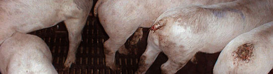
Weaned pigs
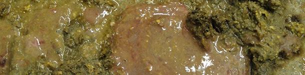
Weaning diarrhea
 |
(c) The Pig Site
|
Because of the stress, many pigs get diarrhea within a week of weaning. It slows down their growth, but can even be lethal. Commonly the diarrhea is caused by E. coli, in which case it may be treated with antibiotics. The medicine is added to the pig feed. In addition, electrolytes can be mixed to the drinking water, decreasing the risk of dehydration. The antibiotic treatment should last 5-7 days.
There is no preventive medication, but by reducing the stress of the weaning may have a huge impact. If at all possible, farrows should not be split in weaning. The best way is to leave the farrow to the pen they were born in, and simply lead the sow away. Good hygiene, warmth and lack of draft are essential at the time of weaning.
Swine dysentery
Swine dysentery is one of the most economically damaging diseases a
farm can experience in terms of medication, mortality, non-marketable
pigs and extra feed costs (
The pig site)
. Swine dysentery is caused by the bacteria
Brachyspira hyodysenteriae, which causes a severe infection in the large intestine of pigs weighing 15-70 kg (can infect also adult pigs). First pigs are usually infected by rat faeces, since rats are hosts for
B.
hyodysenteriae. The symptoms of dysentery are loss of weight, loss of appetite and wet and bloody faeces. The bacteria is spread with the feaces, so it spreads quite easily, and the bacteria survives in the piggery for a long time.
Mortality in pig dysentery is high.
To get rid of the bacteria in the piggery, the whole piggery (or at least the sections with the infected animals) has to be emptied, washed, disinfected and kept empty for two weeks. If all the animals cannot be removed, they must be given preventive medication. All manure handling and cleaning operations must be revieved, and rats eradicated from the building. Boots must be cleaned when moving between sections in the piggery, and each section must have its own tools (such as dung shovels) to prevent any bacteria from spreading between sections.
Swine dysentery in
The Pig Site.
Brachyspira pilosicoli
Br. pilosicoli is a bacteria, which causes diarrhea to pigs a few weeks after weaning. The faeces have no blood, since the bacteria does not destroy the intestines, but the dung resembles cement (gray and lumpy).
Br. pilosicoli infection decreases growth, but it can be treated with antibiotics. Mortality is lower than in swine dysentery.
It is recommended to take faeces samples if
Br. pilosicoli infection is suspected. Since the bacteria dies soon when outside the animal, the sample must be taken directly from the rectum and transferred to a laboratory in a suitable culture medium.
Lawsonia intracellularis
Lawsonia is a bacteria which proliferates in the epithelial cells of the small intestine. It causes decreased growth and occassional diarrhea, which usually start two weeks after weaning. In more serious cases Lawsonia can perforate the intestinal wall, and leading into severe peritoneal infection. Mortality in Lawsonia is low, and it can be treated by mixing antibiotics to the feed. However, the infected pigs continue to spread the bacteria up to 10 weeks, so hygiene is again immensely important.
Lawsonia has a gangrenous form, which can be very severe. It causes constant, bloody diarrhea, and may cause sudden deaths. In post-mortem examinations swollen intestinal lymph nodules and thickened intestinal walls are common findings.
Pig edema disease
The pig edema disease is caused by
Escherichia
coli
enterotoxemia. It infects pigs 1-2 weeks after weaning, and usually attacks the best grown pigs. The bacteria is first ingested by the pig, and soon the bacteria starts to create strong toxins in the guts. The toxins are absorbed and spread into the internal organs,
killing the pig within a day. The condition is described as "in the evening the pigs shiver, in the morning they're dead."
Althought the edema disease cannot be treated, it can be prevented. Before weaning the pigs must be familiarized with high-quality feed, and the amount of feed can be limited during weaning. Adding citric acid or lactic acid bacteria to the feed helps the pigs' digestion and supports the functions of the stomach.
Pig edema disease in
The Merck Veterinary Manual
 |
| Gestation crates, where sows may be kept for weeks. (c) Wikipedia |
Diseases of adult pigs
Leg and hoof injuries
Injuries in the hoof can be caused by accidents, overgrowth of the hoof, a foreign object stuck in the hoof or unfastening of the keratinous layer of the hoof. The hoof may become infected, often because of the bacteria
Fusobacterium necrophorum or
Arcanobacterium pyogenes. These both cause hoof infections also for cows. Due to the rather short lifespan of pigs to be slaughtered, hoof problems rarely cause severe problems. When needed, pig hooves can be trimmed just like cows' or horses'.
Infective arithritis in pigs is often caused by streptococci, which access the joints through open wounds. Arithritis caused by other than Mycoplasma bacteria can be treated with penicillin injections.One of the causing bacteria, E
rysipelothrix rhusiopathie, can cause
swine erysipelasis. This disease is zoonosis, so
it can affect humans. Swine erysipelasis causes clearly visible and elevated red markings in the skin and high fever.
Leg weakness or osteochondrosis (OCD) in pigs affects the surfaces of joints, causing pain when the animal moves. It is usually seen in pigs nearly ready for slaughter, even though the symptoms have developed for a long time. Osteochondrosis is partly hereditary, but strong feeding and fast growth add to the risk of getting ill. OCD can be prevented with selective breeding, soft litter in the pens, spacious pens and calm animal handling to avoid injuries. More about OCD in
The Pig Site.
Tail biting
Tail biting is a very wide-spread and serious problem in piggeries. It is not a disease but a severe behavioral problem. This condition is so common that some producers consider is normal, while actually tail biting is a clear indication of bad animal welfare.
Tail biting means that the animals bite and chew on each others' tails. The tails are often bitten off entirely, and sometimes even the exposed spine is chewed on. This causes severe pain, and is almost impossible to stop once it becomes a habit for the animals. Several factors can induce tail biting:
- Lack of litter and stimuli (toys, chewable objects etc)
- Lack of space to avoid other animals
- Lack of space for proper social behavior
- Bad air quality
- Mixing of animal groups, disturbing their social structure
- Limited feeding or too small feeding trays, not enabling the whole group to eat and drink at the same time
- Lack of minerals or salt in the diet
- Illnesses and parasites
- Stress
Usually a small female pig starts biting the flanks of castrated boars. If the environmental conditions are not improved, more pigs pick up the behavior, and start biting the tails of others. Bitten tails are open wounds, which are prone to infection and when bleeding, encourage the pigs (omnivores as they are) to bite more. Once this becomes a habit, the pigs will not stop it even if they were transferred to ideal conditions. On the contrary, if moved to another group, the biters will only teach the habit to the new animals.
The only way to treat tail biting is to prevent it by good animal handling practices and humane animal husbandry. Docking the tails of piglets or using red lights to avoid the pigs from seeing blood are ineffective in treating the actual cause. They also make the situation worse, when the pigs cannot ease their stress even by tail biting.


